 Catalog
GD1308002
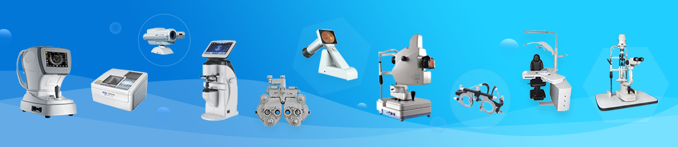
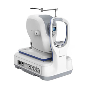
Optical Coherence Tomographer
Model No.: OSE-2800
Overview:
•12mm wide scan for posterior segment
•Deep Choroidal Imaging (DCI) mode reveals more details of the choroid
•3mm Scan Depth
•Comprehensive software analysis for retina, glaucoma and cornea
•16mm Angle-to-Angle Scan
Features:
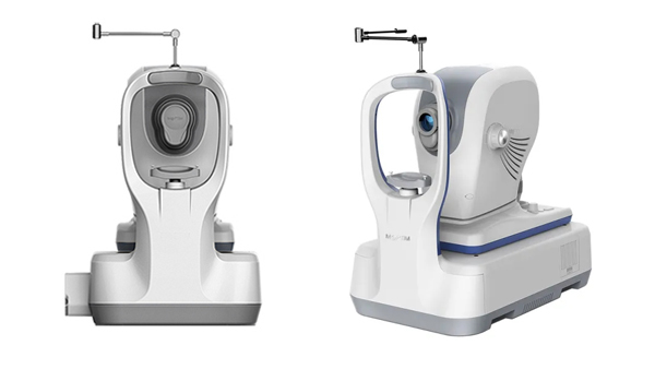
High quality OCT image
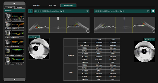
16mm Angle-to-Angle analysis
16mm angle-to-angle anterior scan with data analysis
Epithelial thickness analysis
Provides 6mm diameter cornea epithelium thickness map, which is an important part of diagnostics in
refractive surgery, with many important clinical applications.
Comprehensive software analysis and free upgrade
The OSE-2800 system provides 8 scan patterns to help you improve diagnostic efficiency:
Macular: HD line scan (6mm or 12mm), Cube scan(6mm x 6mm),Six-line radial scan,Multi(X-Y: 5x5)
Disc: Cube scan (6 mm x 6 mm)
Anterior: HD line scan (6 mm), Angle-to-Angle scan (16mm), 6-line radial scan
Specifications:
|
OCT IMAGING |
|
|
Methodology |
Spectral domain OCT |
|
Optical source |
840nm (Center Wavelength) |
|
Axial resolution (optical) |
5 microns (optical), 2.7 microns (digital) |
|
Transverse resolution |
15 microns (optical), 3 microns (digital) |
|
A-scan depth |
3.0 mm |
|
Diopter range |
- 20 to + 20 diopters |
|
Scan patterns |
Macular: HD line scan (6 mm or 12 mm), Cube scan (6 mm x 6 mm), 6-line radial scan, Multi (X-Y: 5 x 5) Disc: Cube scan (6 mm x 6 mm) Anterior: HD line scan (6 mm), Angle-to-Angle scan (16mm), 6-line radial scan |
|
FUNDUS IMAGING |
|
|
Methodology |
IR, en face |
|
Field of view |
40° x 30° |
|
SOFTWARE ANALYSIS |
|
|
Macular thickness analysis |
3D view; En-face analysis; Deep Choroidal Imaging function |
|
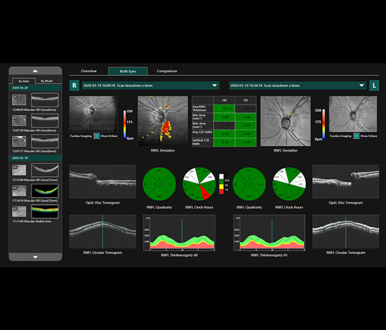
Glaucoma |
RNFL analysis; Ganglion cell analysis; Cup-disk analysis; progression analysis, comparison analysis |
- Copyright 2012 © Yiwu Goddon Vision Technology Co., Ltd. All rights reserved.
No. 12-3, Dongfang Building, Choucheng Street, Yiwu, Zhejiang, China
Tel: +86 0579 85609259 Fax: +86 021 51686909 E-mail: info@goddonoptics.com